Ubiquitin Beyond the Proteome
NoPro-clipping converts ubiquitinated non-protein substrates into peptide-like analytes for LC-MS/MS detection
A new mass spectrometry workflow has made non-protein ubiquitination detectable in cells and tissues, revealing ubiquitin-modified glycogen and small metabolites beyond the protein substrates usually associated with the signaling system.
Called non-protein ubiquitin clipping, or NoPro-clipping, the method targets ubiquitinated substrates that conventional proteomic approaches are likely to miss. Bacterial ubiquitin clippases cut ubiquitin internally, leaving its C-terminal Gly-Gly remnant attached to the non-protein substrate. Sortase-mediated peptide labelling then converts those Gly-Gly-modified molecules into enriched, peptide-like analytes for LC–MS/MS detection.
After validation with in vitro ubiquitinated sugars and glycogen, the team applied the workflow to mammalian cells and tissues. Targeted NoPro-clipping detected ubiquitinated glycogen in glycogen-containing mouse tissues, with the strongest signals in liver and skeletal muscle. In mouse liver, glycogen depletion during fasting coincided with increased glycogen ubiquitination after six hours, followed by loss of signal during longer fasting as glycogen levels fell further.
NoPro-clipping was then used as both a targeted and untargeted readout. In cell and tissue experiments, it tracked glycogen ubiquitination during lysosomal inhibition and glycogen storage disease–relevant perturbations, with isotope-labelled standards supporting quantification in mouse liver. In untargeted mode, the workflow filtered MS features by their dependence on ubiquitin clipping and sortase labelling, recovering glycogen-derived species and identifying endogenous ubiquitinated glycerol and spermine.
The authors suggest that NoPro-clipping could help track this overlooked layer of ubiquitin signalling across tissues, metabolic states, and disease models.
Cryo-EM Meets Chemical Mapping
Focused ion beam SIMS adds chemical identity to cryo-EM images without disrupting vitrified specimens
A correlative cryogenic imaging workflow combines cryo-electron microscopy with focused ion beam secondary ion mass spectrometry to map molecular and elemental signatures inside cells.
The approach addresses a familiar gap in cryo-EM: high-resolution cellular structures can be visualized in near-native specimens, but their chemical identity is often difficult to assign directly. Here, vitrified samples are first imaged by cryo-EM, then transferred under cryogenic conditions to a FIB-SIMS instrument, where a focused gallium ion beam releases secondary ions for time-of-flight mass spectrometry and chemical mapping.
The team validated the approach with labeled bacterial cells before moving to untagged Caulobacter crescentus. Cryo-FIB-SIMS mapped ions including potassium, magnesium, sodium, and phosphate, while cryo-EM localized those signals to features such as storage granules. Compatibility with cryogenic light microscopy and FIB-milled lamellae also extended the workflow toward thicker and eukaryotic specimens.
As a biological test case, the team used the workflow to study uptake of bisphenol-AF, a fluorinated pollutant, by C. crescentus. Bulk measurements showed intracellular accumulation, while proteomics revealed upregulation of efflux-associated proteins, suggesting that the cells were responding to the pollutant but not fully clearing it.
Correlated cryo-EM-FIB-SIMS then localized the bisphenol-AF fluorine signal to cytosolic storage granules rather than diffusely across the cytosol. The result suggests that the bacteria sequester the pollutant in intracellular aggregates, where it may evade removal despite activation of efflux machinery.
More broadly, the workflow offers a way to map chemical signals onto subcellular structure in untagged, near-native specimens, including studies of drug uptake, pollutant accumulation, and molecular localization in more complex cellular samples.
Antidepressants Downstream
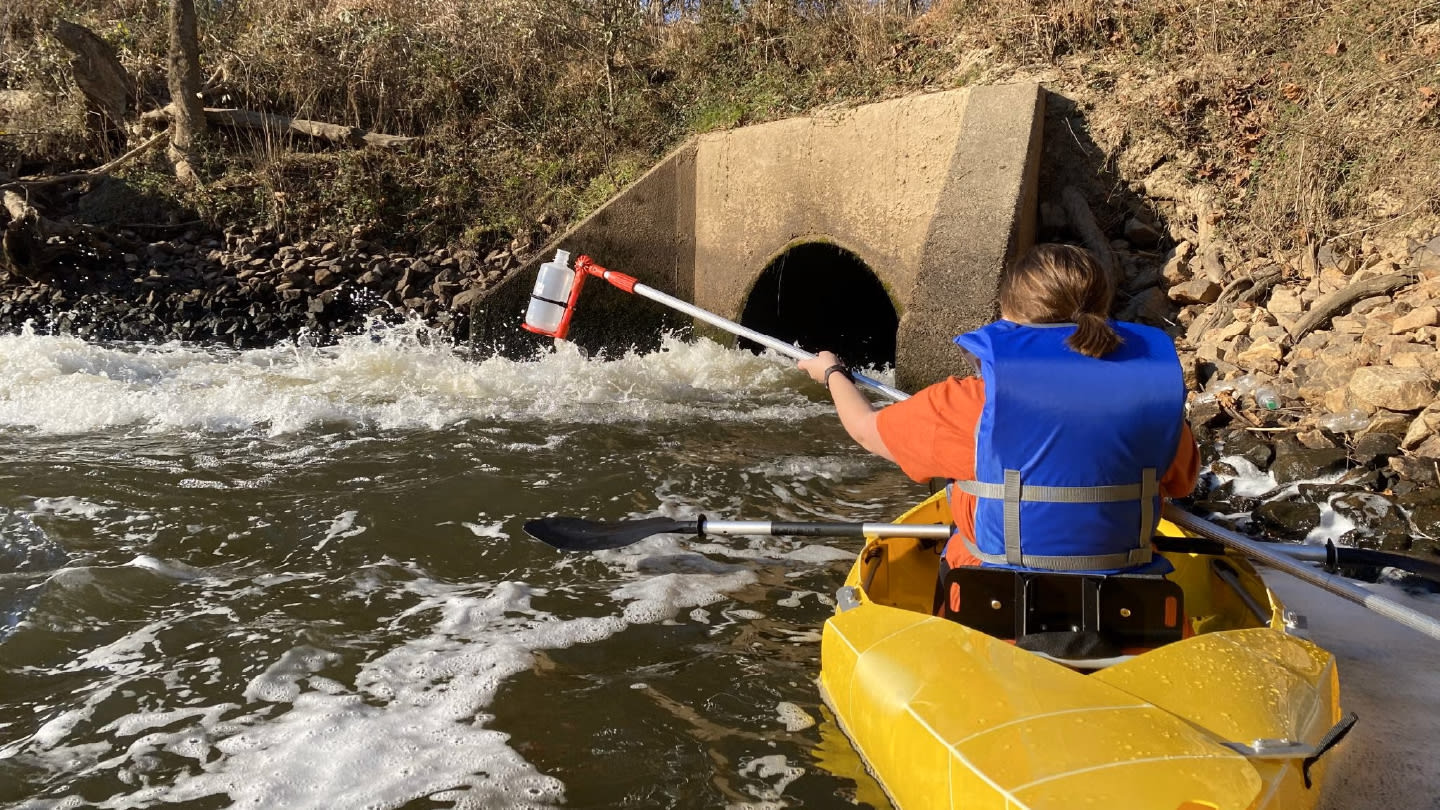
A multidimensional LC-IMS-MS workflow detects antidepressants and metabolites in wastewater-impacted North Carolina waterways
Antidepressants and their metabolites have been detected in wastewater-impacted North Carolina waterways at concentrations that, for some compounds, exceed levels previously linked to behavioral effects in aquatic organisms.
The study, led by Erin Baker’s group at the University of North Carolina at Chapel Hill, developed a multidimensional workflow for monitoring antidepressants in complex environmental samples. Liquid chromatography, drift tube ion mobility spectrometry, collision-induced dissociation, and mass spectrometry were combined to identify and quantify 26 parent antidepressants and eight metabolites, with chiral chromatography used for selected enantiomeric compounds.
“Findings from this study, along with others, reveal that pharmaceuticals are increasingly detected in waterways across the globe, especially near wastewater treatment plant discharge sites, posing a mounting environmental concern,” said Baker in the team’s press release.
The team first built a reference library containing retention times, collision cross section values, fragmentation data, and mass-to-charge ratios for antidepressant standards, with chiral analysis adding information on selected enantiomers that can differ in biological activity. That added confidence was important in wastewater matrices, where structurally similar drugs and metabolites can be difficult to distinguish.
Applied to five sites in central North Carolina, the workflow detected 17 antidepressant-related chemicals. No antidepressants were detected in the lake or in upstream controls at three of the four discharge-impacted sites, while downstream samples contained mixtures of antidepressants, metabolites, and propranolol.
“Studies on effective remediation strategies for these pharmaceuticals are urgently needed to eliminate them from wastewater and mitigate this escalating challenge,” added Baker. “Future research should prioritize broadening sampling efforts across global waterways to fully capture the scope of the issue.”
Tracing Pain Across the Brain
Imaging mass spectrometry and behavioral experiments connect LPA, microglial activation, and prostaglandin signaling after stroke
Imaging mass spectrometry has helped trace how a lipid mediator may drive mirror-image pain after stroke, a rare form of post-stroke pain that spreads to the same side of the body as the brain lesion.
The study, from Kyoto University, focused on lysophosphatidic acid (LPA), a bioactive lipid previously linked to chronic pain. Using desorption electrospray ionization imaging mass spectrometry (DESI-IMS), the team mapped LPA and inflammatory prostaglandins in mouse brain sections after ischemia-reperfusion injury. The measurements showed increased 18:1-LPA in the ischemic core, along the corpus callosum, and in the contralateral cortex.
“The central question underlying our study is: why does pain occur after a stroke, and why does it sometimes spread to both sides of the body?” said first author Hiroyuki Neyama in a press release.
The team then tested that spatial pattern with behavioral experiments, immunostaining, pharmacological inhibition, and local brain injections. Blocking autotaxin reduced LPA signals and bilateral hyperalgesia, while minocycline reduced microglial activation and abnormal pain behavior. Together, the experiments connect LPA production near the ischemic core with inflammatory signaling through the corpus callosum, microglial activation, and downstream prostaglandin signaling in contralateral pain-related regions.
“What impressed us most was our ability to visualize previously unseen inflammatory processes in the brain as molecular images of LPA and PGE₂,” said corresponding author Yuki Sugiura.
The results point to LPA production and microglial activation as possible targets for post-stroke pain, while raising the question of whether similar inflammatory routes contribute to other chronic pain states.
(Mass) Spectacular and Strange
A Biochemical Yawn
A bad night’s sleep may be written all over your face, but a new study suggests it can also leave a “sleepiness fingerprint” in your saliva.
In a randomized crossover study from the University of Zurich, 20 healthy young men completed three conditions: a normal night’s sleep, four nights of moderate sleep restriction, and one night without sleep. Oral fluid samples were then analyzed by liquid chromatography–high-resolution mass spectrometry, with machine learning used to classify the metabolic patterns.
“Until now, sleep deprivation has been impossible to measure biochemically – and yet it is one of the greatest burdens of our time,” said Thomas Kraemer, corresponding author of the study.
The clearest signal came after a full night without sleep. A model based on 12 molecular features correctly identified sleep-deprived samples 94 percent of the time when making a positive classification, while moderate sleep restriction did not produce the same exploitable fingerprint.
For now, the study remains exploratory, though the team is now expanding the work to more than 1,000 samples from shift workers, women, and frequent drivers.
Newsletters
Receive the latest analytical science news, personalities, education, and career development – weekly to your inbox.


