The problem
Autism spectrum disorder (ASD) diagnoses are steadily increasing – yet our ability to detect the condition early has not kept pace. Current diagnostic tools rely on observing behavioural symptoms that emerge years after the first molecular disruptions, by which point valuable opportunities for early intervention may have been lost. Existing methods struggle to detect the faint chemical signs of autism quickly, sensitively, and in a way that is practical for routine clinical use. This diagnostic blind spot is what our work seeks to close.
The solution
As a postdoc at University of Massachusetts Lowell, I studied Alzheimer’s disease and a puzzling chronic syndrome, Gulf War Illness, both of which appear to have some link to dyshomeostasis (that is, imbalance) of metals in the human body. Autism also seems to be accompanied by alterations of the natural abundance of essential metals such as magnesium, copper, and zinc, and anomalous accumulation of metallic neurotoxicants such as mercury, lead, and chromium. (Indeed, some researchers have suggested that the rise in autism diagnoses could be partially explained by environmental exposure to neurotoxic metals.) But it wasn’t until a colleague from my current institution – Andrea De Giacomo, a professor of child neuropsychiatry who studies neurodevelopmental disorders – reached out to ask if our laser-induced breakdown spectroscopy (LIBS) technique could be used to study autism that we then began a project to study Enhanced Laser spectroscopy Techniques for autism Diagnosis in children (ELATED).
Metallomic profiles – a sort of “map” of the abundance of metals in a biological sample – are key to what we do. Basically, they tell us which metals are contained in the sample and define ranges for the amount of each metal that can be considered healthy or pathological.
Many diseases or disorders are accompanied by alterations of these abundances (what earlier I called “dyshomeostasis”). Therefore, by comparing samples from patients suffering from a given medical condition (in our case, ASD) to healthy controls, we can discover if there are systematic differences in their metallomic profiles and associate an altered metallomic profile with the medical condition under investigation.
This is important for various reasons. Many neurological diseases are diagnosed only after symptoms appear – often at advanced stages – so identifying early metallomic changes could enable earlier and more accurate detection. Moreover, different diseases can manifest with very similar symptoms, which could lead to wrong diagnoses and, consequently, ineffective treatments. If, with a simple blood or urine test, we could detect anomalies in the metallomic profiles that we have previously associated with specific conditions, this would provide us with an objective and unbiased instrumental technique to associate with clinical diagnoses. The synergy of the two would be an incredibly powerful tool that, for example, could allow us to diagnose ASD before symptoms even appear, and therefore start therapies or seek more detailed diagnoses at an earlier stage. Or, in the presence of symptoms, we could provide information useful for a differential diagnosis, helping to distinguish between different neurological disorders that may have similar symptoms but are not the same, and could be accompanied by different metallomic profile alterations. If, finally, we could make the analysis virtually noninvasive (for example, by using tiny amounts of blood such as those used in devices to measure blood sugar) and portable, this would mean that patients in underserved or remote areas could have access to timely diagnoses and therapies without having to reach specialized hospitals.
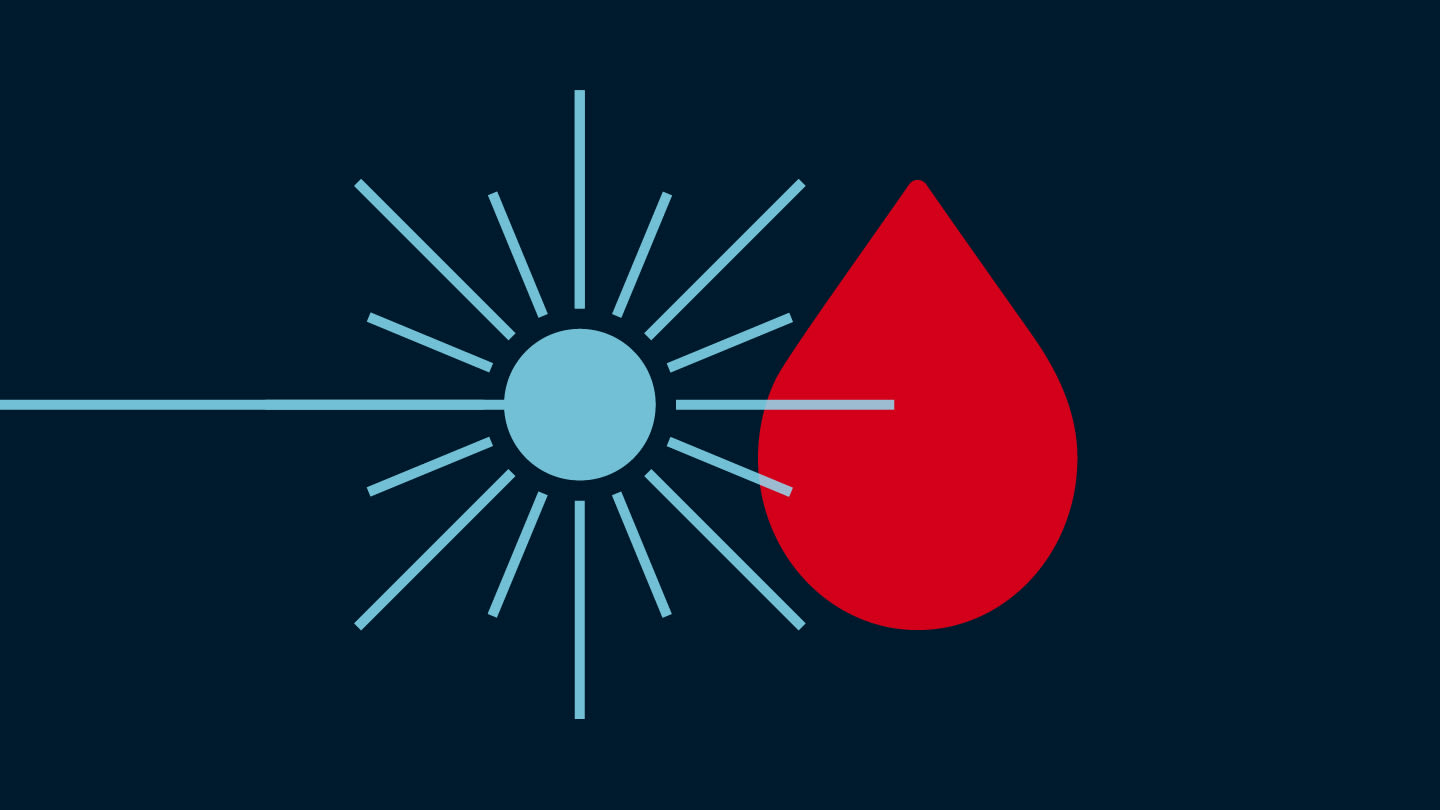
LIBS ticks several of these boxes – and, down the line, it has the potential to make a difference for many families. But it all starts with identifying which metals are in higher (or lower) amounts than they should, and correlating these metallomic profiles with the disorder under investigation.
Nonetheless, there are some practical challenges when analyzing biological fluids.
The first is that working with liquids is not the easiest task with LIBS, because focusing a powerful laser on a liquid tends to cause all sorts of experimental issues, such as sample splashing and quick quenching of the emission signals. To avoid this, we deposit and dry drops of samples on solid substrates prior to interrogating them with the laser (an approach often referred to as surface-enhanced LIBS). This not only takes care of the splashing issues, but, if the substrate is chosen carefully so that it can absorb the incoming laser beam as efficiently as possible, it also helps obtain a more intense signal, which in turn can make the analysis more sensitive.
The second challenge is precisely that – making the analysis as sensitive as possible. Intensifying the LIBS signal is an active area of research, and several groups around the world are testing many different solutions, going from multiple-pulse approaches to adding electric and magnetic fields to make the signal longer-lasting and more intense. The solution that our group proposed several years ago now (back then I was a postdoc at University of Bari) is depositing and drying a drop of a nanoparticle solution on the substrate prior to the analysis. These nanoparticles can act as sort of antennas, essentially “concentrating” the laser power on the sample and consequently intensifying the LIBS signal. This variant of the basic technique, which we called Nanoparticle-Enhanced LIBS (NELIBS) is an effective way to improve LIBS sensitivity. Some fundamental work is still needed before we can fully harness its power for clinical applications, and part of the “ELATED” project is in fact focused on better understanding NELIBS and finding ways to standardize it for routine analysis.
The underlying idea of the ASD project is that making LIBS more sensitive will help us detect smaller traces of elements in patients’ blood, which in turn will make the differences between patients and controls more obvious and the diagnosis more accurate. At the same time, though, we want to do this without adding too much instrumental complexity, such as additional power sources, vacuum systems and so on, because we want to keep the analysis portable, user-friendly, and cheap.
We’re using nanostructured substrates to do this. Our collaborators at University of Catania (Giuseppe Compagnini and his team) are developing a novel method to chemically anchor nanoparticles to the substrates where we will deposit biomedical fluids. This is a sort of NELIBS 2.0, because it enables fine control of the amount, properties, and distribution of the nanoparticles on the substrate. Our initial findings are very encouraging – we obtained significant intensification of the LIBS signal of solutions, exceeding 100 and even approaching three orders of magnitude in some cases. This means that, with this method, we could detect elements with much lower concentration than we would with standard LIBS.
There is another variant of LIBS that we’ve only very recently started studying. Instead of chemically functionalizing substrates with nanoparticles, we are investigating if a similar antenna effect could be produced by generating ordered nanostructures directly on the substrate, using ultrashort lasers. These structures are called Laser-Induced Periodic Surface Structures (LIPSS) – or ripples. Our collaborators from the Italian National Council of Research (CNR), Caterina Gaudiuso and Antonio Santagata, are fabricating and studying various kinds of substrates, using different laser sources and texturization methods to obtain LIPSS with tailored properties to intensify the LIBS signal. It is a challenging, but extremely exciting line of research!
Beyond the solution
Although our pilot study is promising, we need validation of its preliminary results with a much larger patient cohort, and we are currently working on this with our coworkers at the Polyclinic Hospital of Bari. In the future, the patient cohort should also be designed in a way to also allow assessing the effect of common comorbidities (i.e., the presence of other diseases or disorders) that could also affect the signal. Indeed, we are currently conducting such a study!
There are also some engineering issues to be solved before we can move into the clinic. For example, in our research laboratory we look at LIBS spectra and compare them against atomic spectroscopy databases to figure out which elements are responsible for the signals that we observe. For clinical applications, this process would have to be much more streamlined so it could be performed routinely, without requiring highly specialized knowledge for raw data interpretation. The spectroscopic data should be made directly available to clinicians and ready to be turned into medical decisions. And a third aspect would be standardizing the sample preparation for use with a compact and easy-to-use device.
Standardization is another problem that needs addressing. Until relatively recently, and it is still largely the case in research laboratories, LIBS setups were not commercially available compact instruments, but rather homemade systems assembled by combining various blocks, i.e., laser sources, optical elements such as mirrors and lenses, and spectrometers and detectors for signal acquisition. The problem is that, for example, different laser sources (that is, different wavelengths, different pulse amplitudes, etc.) interact differently with matter, and therefore can generate laser-induced plasmas that behave differently (e.g., in terms of different persistence time, emission intensity, degree of ionization, and so on). This, in turn, can profoundly affect the LIBS signals of these different plasmas. Furthermore, with homemade setups, you have a very flexible experimental setup, which means that the number of variables is immense, and different acquisition settings, geometry etc., can make results from different laboratories not always easily and readily comparable. To use the technique in clinical settings, this kind of variability would need to be eliminated, even at the cost of reducing some of our experimental flexibility. The community is moving in this direction, not only thanks to the increasing commercial availability of LIBS instruments, but also by collective research works aimed at improving the interlaboratory comparability of results.
LIBS is obviously far from perfect, but it is clear, talking to and working with biochemists and medical doctors, that it has features for which there is quite a strongly felt need. At its current stage of development, LIBS is already able to meet several of these needs. If we succeed in improving its sensitivity and integrating the intensified LIBS variants into commercially available instruments, especially compact portable ones, I definitely expect this technique to become a staple in biomedical science, as is currently happening in the fields of geochemistry and space exploration – the NASA missions to Mars, for example: both Curiosity and Perseverance rovers include LIBS spectrometers on their mobile lab platforms. The principles of interdisciplinarity and translational science are of great importance to ensure the solutions offered by techniques such as LIBS are brought to the people that need them in the medical community.
The results discussed in this article were presented at the following conferences: 44th Colloquium Spectroscopicum Internationale – CSI 2025 (Ulm, Germany, July 27-31, 2025) and SciX 2025 (Covington, KY, USA, October 5-11, 2025).
The project “Enhanced LAser spectroscopy TEchniques for autism Diagnosis in children (“ELATED”) is funded by the European Union (PRIN-PNRR 2022, CUP H53D23007820001.)
Newsletters
Receive the latest analytical science news, personalities, education, and career development – weekly to your inbox.


