Opening the “Black Box” of Carbon Defects
Atomic-scale modeling and spectroscopy tie broad defect bands in high-temperature carbon materials to specific local structures.
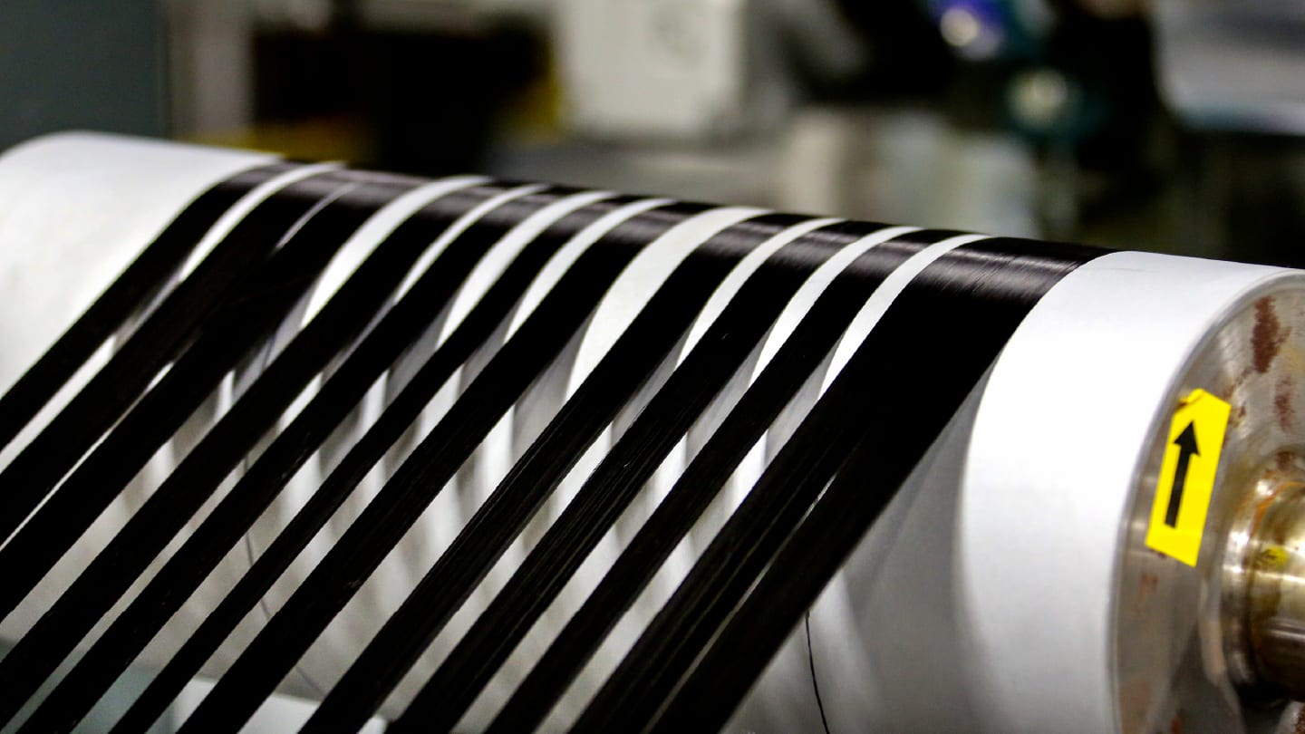
A combined spectroscopy and computational study has clarified the structural origins of disputed defect peaks in carbon fibers and related high-temperature carbon materials.
Researchers at Chiba University examined isotropic pitch-based carbon fiber as a model system, combining experimental Raman, infrared, and XPS measurements with density functional theory calculations. The study compared fibers heated from 773 to 3173 K with 34 graphene models containing oxygen functional groups, vacancy defects, sp³ carbon, and non-hexagonal rings.
The calculations showed that, when charging effects, C–N bonds, and adventitious carbon can be ruled out, the disputed 285 eV XPS feature can instead arise from carbon atoms surrounded by three rings, including at least one heptagon, octagon, or larger vacancy defect. The finding challenges a common shortcut in carbon analysis: assigning that peak directly to sp³-hybridized carbon.
Raman spectra told a similar story of structural overlap. Peaks between roughly 1500 and 1550 cm−1 were traced to C=C stretching in hexagonal rings whose local bonding environment had been distorted by nearby non-hexagonal rings or oxygen-containing groups such as cyclic ethers. In the carbon fibers heated at 1873 K or above, the dominant defect structures were assigned to edges, cyclic ethers, and non-hexagonal rings.
“The exact atomic-level origins of the specific peaks derived from these defects have long remained a ‘black box’ in the field of carbon science,” said study lead Yasuhiro Yamada in the team’s press release. “By finally clarifying how specific defects like non-hexagonal rings and cyclic ethers influence Raman and XPS spectra, we can now evaluate the structures of various carbon materials with unprecedented precision.”
Rather than treating Raman and XPS defect bands as broad disorder markers, the work offers a more explicit route from spectral features to atomic structure in carbon materials processed at high temperatures.
A Raman Cavity for Tiny Gas Samples
An asymmetric hollow-core fiber cavity boosts Raman sensitivity enough to detect multiple gas species from milliliter-scale samples.
A fiber-resonant Raman sensor has detected gaseous and dissolved-phase species from milliliter-scale sample volumes, offering a compact route to simultaneous multi-gas analysis in breath, batteries, and insulating oils.
The system centers on an asymmetric fiber resonant cavity built from hollow-core anti-resonant fiber and mirrored large-mode-area fibers. Raman spectroscopy can identify multiple gas species with a single excitation wavelength, including homonuclear molecules such as H₂ and O₂, but its weak scattering signal limits sensitivity. To address this, the researchers developed a model of Raman signal generation inside the fiber resonant cavity and used it to balance intracavity laser build-up against Raman signal loss.
That balance proved important. A more reflective input mirror increased the circulating laser power, but it also made it harder for Raman photons to leave the cavity. By making the input mirror slightly less reflective than the output mirror, the researchers increased the Raman signal 170-fold compared with hollow-core fiber alone and 36-fold compared with a geometry resonant cavity. The reported limit of detection reached 0.01 ppm·bar, with gas consumption between 0.3 and 2 mL.
The team then nested the cavity with a separation membrane, allowing gases to be measured directly or to diffuse from liquid samples into the hollow-core region. In breath samples, the system detected carbon dioxide isotope peaks, methane, and hydrogen. In overcharged battery electrolyte, it detected dissolved hydrogen, carbon dioxide, methane, ethylene, carbon monoxide, and hydrofluoric acid, and it also tracked fault-related gases released into mineral insulating oil after electrical breakdown.
The authors suggest that lower-loss hollow-core fiber could push sensitivity further, while the coaxial membrane-cavity design may also be adaptable to other scattering- and absorption-based spectroscopic techniques.
Brightness Without the Blur
A hafnium chloride scintillator screen pairs strong radioluminescence with micropore-guided light confinement to improve both brightness and sharpness.
Indirect X-ray imaging often forces scintillator screens into a compromise: make them thick enough to capture more X-rays, and the extra light output comes at the cost of lateral scattering and blurred images. A hafnium chloride scintillator screen now addresses both sides of that trade-off, pairing high radioluminescence with a micropore architecture that limits optical crosstalk.
The material component was built around hafnium-based organic–inorganic metal halides whose light emission could be tuned by changing the length of the organic cation. One compound, TMA₂HfCl₆, produced the strongest ultraviolet-excited emission. Inductively coupled plasma mass spectrometry showed that zirconium was present only at trace levels, helping rule out impurity-driven luminescence.
A set of spectroscopic measurements pointed to thermally activated delayed fluorescence as the source of the high light output. The key emission band grew stronger as temperature increased, and temperature-dependent photoluminescence lifetimes supported the conversion of triplet excitons into emissive singlet states. Excitation-power-dependent photoluminescence, Raman spectra, thermoluminescence measurements, and density functional theory calculations further tied the emission to charge transfer within distorted hafnium chloride units.
The resulting scintillator reached a reported light yield of 56,563 ± 1,250 photons/MeV and a detection limit of 23.86 nGyair/s, while retaining most of its radioluminescence under repeated X-ray exposure. To keep that strong output from blurring the image, the team loaded the material into silicon micropore arrays. The best-performing screen used 25 μm pores with a 95 percent filling factor and reached 31.41 lp/mm at a modulation transfer function of 0.2, outperforming commercial gadolinium oxysulfide and thallium-doped cesium iodide screens.
By linking exciton harvesting with light management, the study suggests a route to scintillator screens that are both bright and spatially precise.
Proton Transfer in Motion
The study moves beyond a one-step proton-jump picture by resolving skeletal motions that participate during and after transfer.
A multidimensional spectroscopy experiment has captured the coupled electronic and vibrational motions that drive ultrafast proton transfer in 10-hydroxybenzo[h]quinoline, a model system for excited-state intramolecular proton transfer.
The University of Washington-led team used two- and three-dimensional electronic–vibrational analysis to follow the molecule after near-ultraviolet excitation. Broadband near-ultraviolet pump pulses initiated the reaction, while a mid-infrared probe tracked fingerprint-region vibrations between 1300 and 1550 cm−1. By correlating electronic excitation frequencies with vibrational signatures, the approach resolved structural motions that conventional transient absorption or fluorescence measurements could only infer.
The spectra showed that the proton transfer was not a simple one-step jump. As the molecule changed from its photoexcited enol form to the keto product, electronic and vibrational motion remained linked through a high-frequency vibration near 1375 cm−1. The proton transfer and loss of this coherent motion both occurred in roughly 15 fs, supporting a non-adiabatic mechanism in which electronic and nuclear motion evolve together.
The three-dimensional analysis added the lower-frequency part of the picture. Vibrations near 550 and 770 cm−1 were coupled to the main proton-transfer coordinate, suggesting that slower skeletal motions participate during the reaction. Other modes between 200 and 1334 cm−1 appeared after the transfer, consistent with vibrational energy spreading through the molecule as the excited product relaxed.
By separating motions that help drive proton transfer from those that follow as the molecule relaxes, the study gives future simulations a more detailed benchmark for modeling non-adiabatic chemistry. The same electronic–vibrational approach could now be used to test how electronic and nuclear motion work together in other fast photochemical processes, from photoisomerization to charge and energy transfer.
A New Engine for Vibrational Spectroscopy
The method delivers large speed gains while retaining spectral features that static harmonic calculations often miss.
A deep learning molecular dynamics framework has simulated infrared and Raman spectra across molecules, crystals, aggregates, and peptides, offering a faster route to vibrational spectra that retain more physical detail than harmonic calculations.
The approach combines DetaNet, a tensor-aware neural network, with molecular dynamics to predict energies, forces, dipole moments, and polarizabilities along simulated trajectories. Those time-dependent dipole and polarizability signals are then converted into infrared and Raman spectra. Unlike conventional harmonic calculations, the dynamic approach can capture anharmonicity, thermal fluctuations, and vibrational coupling; unlike ab initio molecular dynamics, it avoids recalculating the electronic structure at every step.
The researchers trained the model on the QMe14S dataset, which contains more than 186,000 small organic molecules with energies, forces, dipole moments, and polarizabilities. Tests on isolated molecules showed that DetaNet-based molecular dynamics improved agreement with experimental infrared and Raman spectra compared with harmonic quantum chemistry, especially in high-frequency stretching regions. Adding thermostated ring polymer molecular dynamics, which accounts for nuclear quantum effects, further improved peak positions and spectral shapes.
The framework was then extended to more complex systems using limited transfer learning. Fine-tuning on around 2,000 system-specific configurations was enough to reproduce spectra for paracetamol and silicon dioxide crystals, molecular aggregates, and a representative pentapeptide with good agreement against experiment or ab initio reference data.
The speed advantage was substantial. DetaNet molecular dynamics achieved more than a thousand-fold speedup over Gaussian-based ab initio molecular dynamics for isolated molecules, and roughly 750- to 1,000-fold speedups for larger crystals, aggregates, and peptide systems.
While the authors describe the method as foundational rather than fully universal, they argue that it could support faster vibrational spectral assignment and molecular structure recognition across broader chemical systems.
Newsletters
Receive the latest analytical science news, personalities, education, and career development – weekly to your inbox.