Identifying where carbon–carbon double bonds fall within fatty acyl chains has remained a blind spot in lipidomics, limiting our understanding of how subtle structural variations drive biological processes. Conventional approaches can localize these C=C sites, but only with specialized reagents or custom instrumentation that compromise sensitivity and accessibility – limiting their use in most lipidomics labs.
In a new Nature Communications study, researchers present LC=CL, a computational tool that extracts C=C positional information directly from standard reversed-phase LC–MS/MS datasets. By leveraging retention time profiles of more than 2,400 experimentally verified ω-resolved lipid species, LC=CL uses machine learning to map and assign double-bond positions automatically, without altering routine workflows. Benchmarking against established techniques such as electron-activated dissociation and Paternò–Büchi derivatization, the approach not only matched their accuracy but identified significantly more species from human plasma samples.
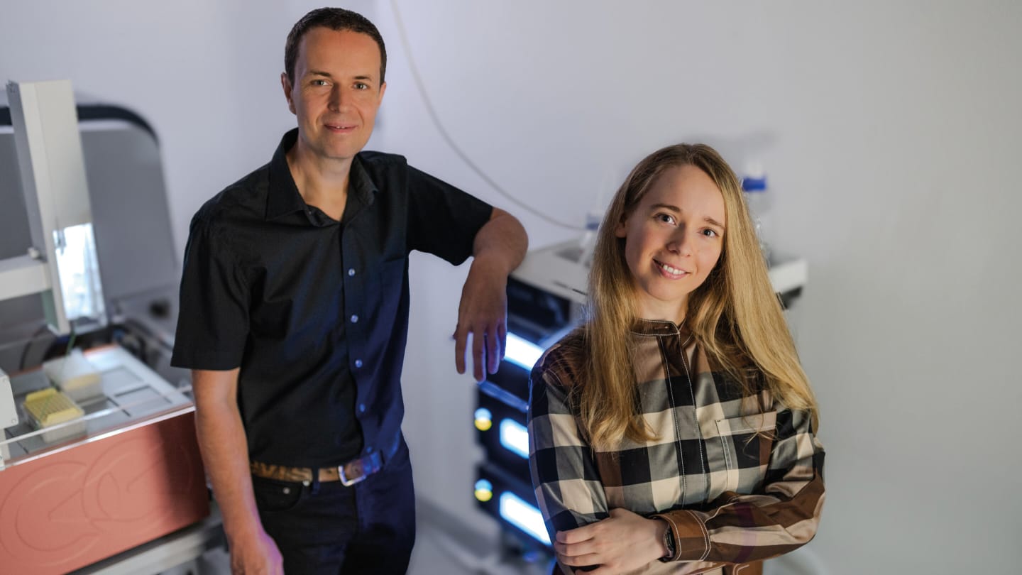
To explore how this method works, the challenges it overcomes, and the issues it could help to address, we reached out to Leonida Lamp and Jürgen Hartler, first and last authors of the study, to find out more.
For readers less familiar with lipidomics, could you briefly explain what omega positions are in fatty acids – and why they matter for health and disease?
In fatty acids, the omega position describes where the first double bond is located counting from the methyl end – the “omega” or final carbon of the chain, in other words. Since free fatty acids can be harmful in the body, they’re usually incorporated into complex lipid molecules. Importantly, even in these complex lipids, the methyl end of each fatty acyl chain is still chemically defined as an omega end. Thus, when we speak about omega-3 or omega-6, we generally refer to lipids with their first double bond three or six carbons away from a chain’s terminal methyl group.
A key reason why this matters is because our bodies cannot make the essential omega-3 and omega-6 fatty acids on their own – so if we don’t eat them, we risk serious health consequences. Without these building blocks, the body tries to improvise by producing a “fallback” omega-9 fatty acid called mead acid. While this helps to keep cells functioning, it also signals a state of deficiency, which can alter inflammation, metabolism, and brain function.
Our newly developed method revealed that even cPLA2, a key enzyme in inflammatory events, can replace its usual omega-6 substrate with a complex lipid containing mead acid. This shifts the balance of inflammatory responses in ways that are not yet fully understood, and may contribute to disease. As growing evidence links omega positions not only to inflammation but also neurological disorders, cancer, and metabolic conditions, understanding these positions may prove crucial for both prevention and therapy.
Could you describe, in a nutshell, how your method works, and how it enables the routine identification of omega positions?
We discovered that our reversed-phase chromatographic setup can clearly separate complex lipids with different omega positions. To take advantage of this, we developed a strategy in which lipids are generated in a cell culture with stable-isotope labels marking their omega position. However, routine users of our method don’t need isotope labels. We consolidated the results into a database that now contains the elution profiles of more than 2,400 omega-position-resolved lipid species.
In practice, the workflow is simple: (i) retention times in the database are calibrated to the specific chromatographic conditions with an algorithm that achieves accuracy to within just a few seconds; (ii) MS/MS fragmentation provides structural evidence for the lipid molecular species, though not their omega positions; (iii) when the measured retention time and lipid identity match a database entry, the omega position can be determined.
In short, the method enables reliable, large-scale identification of omega positions with conventional MS setups.
What motivated your team to develop a new method for pinpointing omega positions? What gaps or limitations in existing approaches were you aiming to overcome?
Existing approaches for pinpointing omega positions require either chemical derivatization or specialized instrumentation, limiting their accessibility. In our publication, we outlined the main barriers currently holding back broader adoption: (i) specialized equipment or chemistry is required, often involving hazardous reagents; (ii) some approaches only work in positive ion mode, even though many lipid classes ionize better in negative ion mode; (iii) the resulting spectra are often too complex for unambiguous structural assignment, especially when multiple unsaturated chains are present; (iv) yields of characteristic ions are frequently low, leading to reduced sensitivity; and (v) quantification is often compromised. To summarize, the field lacked a method that is both straightforward and broadly accessible, yet still accurate, sensitive, and scalable. This gap motivated us to design a new approach that overcomes these barriers and enables routine, large-scale omega-position analysis.
Was there a key breakthrough or “eureka” moment during development?
Our “eureka” moment came when we first realized that reversed-phase chromatography could actually separate lipids by their omega positions. Referring to both the literature and prior experience, we knew that reversed-phase chromatography can, at least partially, separate sn-position isomers – complex lipids that are almost identical, but for where their fatty acids are attached. Such sn-positional differences are typically reflected in fragment ion intensities in MS/MS spectra, and our LDA software was the first tool capable of identifying sn-positions from these fragments.
However, when we analyzed our first samples with the new chromatographic method, we noticed something puzzling: three perfectly baseline-separated peaks for lipids with the same molecular mass. LDA annotated all three peaks as identical, with the same head groups and fatty acids, at the same sn-positions – with MS/MS spectra providing no additional clues. The question then arose: What structural feature could be responsible for this separation?
The breakthrough came when we compared these results to isotopically labeled omega-6 lipids, provided through our collaboration with a group at UCSD studying these lipids in inflammation. Strikingly, for the labeled species corresponding to those three peaks, only a single peak appeared – the first one. Additional experiments including omega-7 and omega-9 species confirmed the answer: the three peaks represented omega-6, omega-7, and omega-9 positional isomers.
What was the greatest technical or analytical challenge you faced – and how did you overcome it?
While the separation of omega-positions was an exciting finding, the real challenge was to determine whether it could be reliably used for automated assignment. This required retention time precision on the order of just a few seconds – beyond what typical experimental setups can consistently achieve across multiple measurement batches. With this in mind, we developed a computational calibration strategy based on cubic spline interpolation. This approach delivered the necessary accuracy, and can even outperform more complex machine-learning methods (including deep neural networks) in similar scenarios. With this high-precision calibration in place, determining omega-positions is now accessible to any lab equipped with an MS/MS-capable mass spectrometer.
What kinds of biological or clinical questions do you think this method could help address?
This is where the real potential lies. Many biological processes depend on the exact omega position of lipids. Let’s start with inflammation: many signaling molecules involved in inflammation derive from specific omega-series fatty acids. With LC=CL, we may uncover previously hidden lipid signatures that sustain chronic inflammation in diseases such as arthritis or inflammatory bowel disease, with further potential as biomarkers for early detection or treatment monitoring.
Furthermore, in cardiovascular disease, epidemiological studies link omega-3-derived molecules to cardioprotection, while many omega-6-derived molecules promote inflammation. Having the ability to precisely distinguish and quantify these lipids in plasma could improve patient risk stratification, and even inform personalized nutritional interventions. More broadly, tracing how dietary fatty acids (from fish oils, seed oils, etc.) are incorporated into complex lipids with omega-positional accuracy could strengthen diet-health connections.
Additionally, in cancer, cells rewire lipid metabolism to fuel growth and invasion. The SCD1 enzyme, for example, produces omega-7 and omega-9 lipids and is strongly linked to tumor aggressiveness and metastasis. Because SCD1 is also critical in normal tissues, targeting it directly for cancer treatment has thus far proved difficult. But by resolving lipids at the omega level, LC=CL could expose vulnerabilities unique to cancer cells that may be exploited for new therapeutic strategies.
What are the next steps for your team? What do you anticipate will be the main barriers to more widespread adoption by the broader lipidomics field?
Looking ahead, the real opportunity lies in scaling and broadening the method. The barriers are low, technically speaking: any lipidomics lab using reversed-phase LC–MS can adopt LC=CL with standard 30- or 60-minute gradients, as already shown by our collaborators in Vienna. The immediate task is to keep expanding our reference database. Currently, it’s mostly made up of even-chain mammalian lipids; adding odd-chain species, plant- and microbial-derived lipids, and oxidized lipids will greatly extend its reach. Importantly, this expansion isn’t limited to our group alone, as any researcher can contribute new entries to the database through their own experiments.
What excites us most, though, are the biological applications. As LC=CL can also work retrospectively, we can revisit existing studies to see whether omega-resolved lipid signatures were hiding in plain sight all along. In parallel, combining LC=CL with functional studies, e.g. in inflammation, cancer, or cardiovascular disease, could reveal exactly how omega positions shape enzyme selectivity and disease pathways.


