Conexiant
Login
  • The Analytical Scientist
  • The Cannabis Scientist
  • The Medicine Maker
  • The Ophthalmologist
  • The Pathologist
  • The Traditional Scientist
The Analytical Scientist
  • Explore

    Explore

    • Latest
    • News & Research
    • Trends & Challenges
    • Keynote Interviews
    • Opinion & Personal Narratives
    • Product Profiles
    • App Notes

    Featured Topics

    • Mass Spectrometry
    • Chromatography
    • Spectroscopy

    Issues

    • Latest Issue
    • Archive
  • Topics

    Techniques & Tools

    • Mass Spectrometry
    • Chromatography
    • Spectroscopy
    • Microscopy
    • Sensors
    • Data & AI

    • View All Topics

    Applications & Fields

    • Clinical
    • Environmental
    • Food, Beverage & Agriculture
    • Pharma & Biopharma
    • Omics
    • Forensics
  • People & Profiles

    People & Profiles

    • Power List
    • Voices in the Community
    • Sitting Down With
    • Authors & Contributors
  • Business & Education

    Business & Education

    • Innovation
    • Business & Entrepreneurship
    • Career Pathways
  • Events
    • Live Events
    • Webinars
  • Multimedia
    • Video
Subscribe
Subscribe

False

The Analytical Scientist / Issues / 2020 / May / On the Origin of Color
Microscopy Spectroscopy

On the Origin of Color

The 170-year-old mystery behind the first color photographs has been solved

By Lauren Robertson 05/06/2020 1 min read

Share

Using a combination of electron microscopy, X-ray analysis and UV-Vis spectroscopy, a team of researchers from the Centre de Recherche sur la Conservation (CRC CNRS-MNHN-Ministère de la Culture) has finally uncovered the origin of the world’s first color photos (1).

Figure 1 – French physicist, Edmond Becquerel’s “Photochromatic images” of the solar spectrum, considered to be the world’s first color photographs. Credit: Musée Nicéphore Niépce, Chalon-sur-Saône, France.

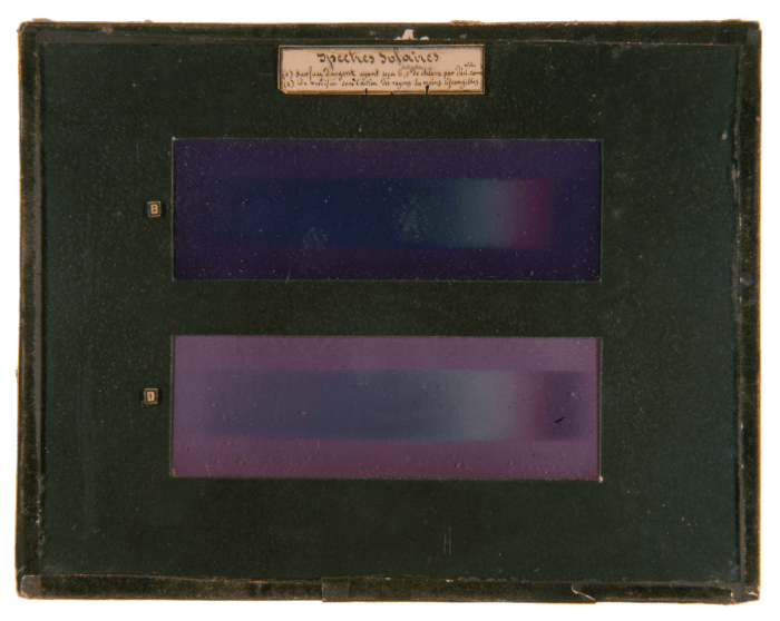
In 1848, French physicist, Edmond Becquerel, produced “photochromatic images” of the solar spectrum in full splendor. Since then, the origin of the colors in these photos has been widely debated within the scientific community. Now, more than 170 years later, we finally have an answer.

Using UV-Vis spectroscopy, the team at CRC observed that visible light absorption by the images could be due to silver nanoparticles of various shapes and sizes. They confirmed their theory by viewing the photochromatic material with electron microscopy, visualizing micrometric silver chloride grains and silver nanoparticles. Transmission electron microscopy was then used to study the size distribution of these nanoparticles, and their location within the silver chloride matrix, which pointed toward a plasmonic origin to the photochromatic colors.

“We propose that silver nanoparticles within the light-sensitized layer of the photographs reorganize when exposed to light – some fragment and others coalesce – enabling it to absorb different colors,” says Victor de Seauve, lead author of the paper. “The color we see is the only one not absorbed – the one that caused this reorganization.” By combining spectrometry with electron microscopy the team were able to detect surface plasmon resonance around silver nanoparticles to confirm the hypothesis.
 

Figure 2 - Silver chloride is reduced in metallic silver nanoparticles under the electron beam in a scanning electron microscope. Credit: Victor de Seauve, Centre de Recherche sur la Conservation.

“The greatest challenge we faced during this study was the sensitivity of silver chloride to the probes of the various analytical techniques employed,” says de Seauve. “By exhaustively studying beam damages, we managed to avoid sample damage during electron microscopy experiments by coating our samples in carbon and working at low temperatures,” he adds. “This is a great step toward understanding how beam damages can affect the analysis of such sensitive materials.”

Newsletters

Receive the latest analytical science news, personalities, education, and career development – weekly to your inbox.

Newsletter Signup Image

References

  1. V de Seauve et al., Angew Chem Int Ed (2020). DOI: 10.1002/ange.202001241

About the Author(s)

Lauren Robertson

By the time I finished my degree in Microbiology I had come to one conclusion – I did not want to work in a lab. Instead, I decided to move to the south of Spain to teach English. After two brilliant years, I realized that I missed science, and what I really enjoyed was communicating scientific ideas – whether that be to four-year-olds or mature professionals. On returning to England I landed a role in science writing and found it combined my passions perfectly. Now at Texere, I get to hone these skills every day by writing about the latest research in an exciting, creative way.

More Articles by Lauren Robertson

False

Advertisement

Recommended

False

False

The Analytical Scientist
Subscribe

About

  • About Us
  • Work at Conexiant Europe
  • Terms and Conditions
  • Privacy Policy
  • Advertise With Us
  • Contact Us

Copyright © 2025 Texere Publishing Limited (trading as Conexiant), with registered number 08113419 whose registered office is at Booths No. 1, Booths Park, Chelford Road, Knutsford, England, WA16 8GS.