Researchers have developed a method to generate elemental maps of nanoparticles and soft/biomaterials in frozen solvent using cryo-transmission electron microscopy (cryo-TEM) combined with electron energy loss spectroscopy (EELS). By carefully managing image alignment and drift between exposures, the team from Tohoku University, Japan, reliably detected and mapped key elements – even in nanoparticles as small as 10 nm.
They demonstrated this by mapping silica, carbon, phosphorus, and calcium in biologically relevant systems such as protein-coated nanoparticles and hydroxyapatite.
“It’s a step forward in being able to visualize not just what a sample looks like under cryo conditions, but what it’s made of – with nanometer-scale resolution,” says Koji Yonekura, Professor of Institute of Multidisciplinary Research for Advanced Materials (IMRAM) and co-authors, Daisuke Unabara, Tasuku Hamaguchi and Yohei Sato, of the study.
The new method could enable advanced analysis of various materials, including biomaterials, medical materials, food, catalysts, and inks.
Yonekura and Hamaguchi come from the field of cryo-EM for structural biology – mainly working on proteins. “But in recent years, we’ve seen growing interest from the materials science side, especially from researchers who want to study soft materials and nanoparticles in their native, hydrated states,” say Hamaguchi and Yonekura. “That made us wonder: if cryo-EM is already powerful enough to visualize proteins at near-atomic resolution with very high sensitivity, why not apply the same principles to elemental mapping?”
The main challenge of elemental mapping in frozen solvents is signal-to-noise – frozen water adds background, and the elements researchers usually care about (such as carbon or phosphorus) produce weak signals.
“We realized that the high-resolution, low-dose techniques we use in biology, combined with careful image correction, could help overcome those limitations,” says Yonekura.
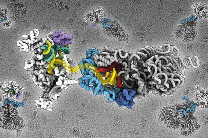
“What surprised us most was that we were able to detect extremely weak core-loss signals from the protein coating – despite its high sensitivity to electron irradiation,” he says. “With careful dose management and signal extraction, we managed to pull out reliable elemental information even under such challenging conditions. This finding really expanded our expectations for what’s possible in cryo-EELS of bio and soft materials.”
The researchers believe that their technique can be immediately applied to liquid-phase bio/soft materials to clarify whether specific elemental components have been successfully introduced, and whether the intended chemical modifications are truly achieved – by combining high-resolution morphological observations with elemental mapping, and doing so without any special treatment.

“We’d like to see many such applications in the near future,” they say. “Being able to directly confirm both structure and composition at the nanoscale would be invaluable for optimizing these materials and understanding their functions.”
The study brought together imaging, spectroscopy, and computational tools – the kind of integration Unabara, Hamaguchi, Sato and Yonekura see as essential for the future of analytical science.
“Analytical challenges today often can’t be solved with a single tool or technique,” they say. “We need hybrid approaches – especially when working at the nanoscale or in complex biological systems. In our case, combining imaging with EELS required careful calibration and computational corrections to get meaningful data. Without image alignment, drift correction, and signal optimization, the maps would’ve been too noisy or inaccurate. So yes, we think the future of analytical science lies in smart integration – where we tailor the method to the question, not the other way around.”


