Antimicrobial resistance (AMR) is often described as one of the greatest global health threats of our time. An estimated 4.95 million deaths were associated with AMR in 2019, and by 2050 AMR is expected to cause 10 million deaths annually. Scientists of all stripes will, undoubtedly, play a part in combatting AMR. But what about analytical scientists?
One area is the need for more rapid antimicrobial sensitivity tests. Culture techniques often require 48 hours to determine an AMR profile, leading to the use of presumptive antibiotic therapies.
In response, researchers from the University of Southampton, UK, have developed a multi-excitation Raman spectroscopy (MX-Raman) method that eliminates the need for sample preparation – working directly on bacterial samples, with no need for culturing or labeling – and uses deep learning to deliver results in minutes.
The researchers believe their platform could screen for bacterial infection and recommend effective antibiotic treatment ahead of confirmation by conventional techniques, improving clinical outcomes and reducing the spread of AMR.
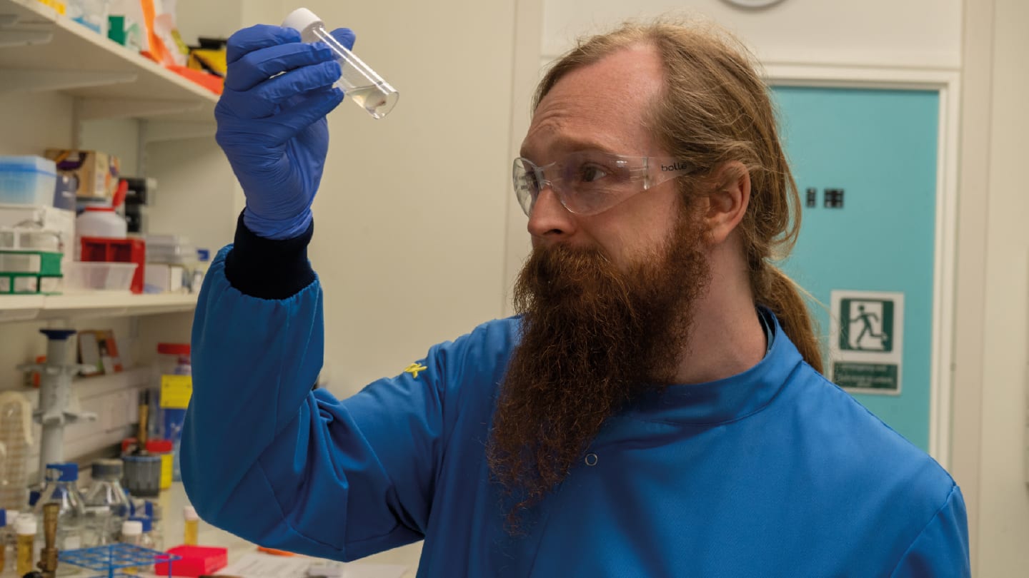
Lead author Callum Highmore tells us more.
Why Raman?
The project developed as there was already a very strong base of Raman spectroscopy at the University of Southampton when I joined the team, and as part of the National Biofilms Innovation Centre we were keen to apply some advanced techniques to microbiological problems. Raman has a lot of strengths as a microbial identification technology, and applying Raman spectroscopy to AMR was an easy choice to make because it is a profoundly important problem that needs all the attention it can get.
Why MX-Raman?
AMR is an area that's recently had a lot of new technology thrown at it and there are some great developments. There are some high-throughput antimicrobial sensitivity tests (AST) that use microfluidic impedance cytometry at the single cell level, like iFast. There have been recent studies on the use of immunodiagnostics in the form of lateral flow assays for AST, and improvements to established techniques such as MALDI-TOF and metagenomics support their use for detecting AMR at scale.
MX-Raman has some way to go before it is a viable diagnostic tool, but I think it has huge potential. It’s fast, where a spectral dataset can be produced in minutes. We’re working on optimizing it for direct analysis of patient samples so that we could entirely cut out the need for bacterial isolation or sample enrichment by culture techniques. MX-Raman is also simpler than other Raman techniques such as surface-enhanced Raman spectroscopy (SERS) in terms of sample processing, so it could be more suitable for clinical translation in that respect.
More generally, Raman is able to give phenotypic information about bacteria, e.g. the presence of biofilms, which isn’t possible for metagenomic approaches, and it can be integrated into AI analyses which gives it an advantage over lateral flow techniques. The limitations include that there are currently fewer and smaller Raman datasets than there are for bacterial genomes, and there is no consensus on standardizing Raman datasets that we can use to build these large-scale models. We also need to work hard to improve the sensitivity of Raman to pull out clinically relevant pathogen data from the background of the host sample.
Did any of your findings surprise you?
In the early stages of the project, it was a real surprise to see how visually different the spectra of different bacteria are, sometimes even at the strain level. It was when we started getting that resolution in our data that it dawned on us that we could push our new MX-Raman methodology all the way into measuring phenotypic differences between similar bacterial populations.
What was the biggest hurdle you had to overcome?
This work has been deeply multidisciplinary. I’m a microbiologist, and I’ve been working with physical chemists and computer scientists to advance this technology towards a medical application. To make this project work, we’ve had to untangle all our preconceptions about each others’ fields, understand some of the intricacies of how these fields can fit together, and find a common language to collaborate effectively. So our biggest challenge has essentially been in dismantling the silos!
How far away is MX-Raman from clinical use?
The biggest barrier to translation is applying this methodology directly to real patient samples, cutting out the need for culture or other sample processing steps entirely. We also still need to build data libraries of bacterial Raman spectra that are large and robust enough to account for the massive heterogeneity found in these clinical samples. We’re already on our way with tackling these issues, and if we manage to keep our momentum up, I’m hoping to be testing MX-Raman alongside existing clinical procedures in the next 5 years.
Next steps?
There are a few things we’re working on across the team to push the whole project forwards. We are optimizing our data processing and analysis pipeline to speed up and standardize our workflows. We have a couple of studies underway looking at how MX-Raman works for different clinical scenarios and other aspects of microbial characterization. I’m focused on applying our methodology to clinical sputum samples to see if we can pull out the same quality of information directly from infected sputum as we can from pure bacterial samples. Preliminary data are looking promising!
Newsletters
Receive the latest analytical science news, personalities, education, and career development – weekly to your inbox.


