Conexiant
Login
  • The Analytical Scientist
  • The Cannabis Scientist
  • The Medicine Maker
  • The Ophthalmologist
  • The Pathologist
  • The Traditional Scientist
The Analytical Scientist
  • Explore

    Explore

    • Latest
    • News & Research
    • Trends & Challenges
    • Keynote Interviews
    • Opinion & Personal Narratives
    • Product Profiles
    • App Notes

    Featured Topics

    • Mass Spectrometry
    • Chromatography
    • Spectroscopy

    Issues

    • Latest Issue
    • Archive
  • Topics

    Techniques & Tools

    • Mass Spectrometry
    • Chromatography
    • Spectroscopy
    • Microscopy
    • Sensors
    • Data and AI

    • View All Topics

    Applications & Fields

    • Clinical
    • Environmental
    • Food, Beverage & Agriculture
    • Pharma and Biopharma
    • Omics
    • Forensics
  • People & Profiles

    People & Profiles

    • Power List
    • Voices in the Community
    • Sitting Down With
    • Authors & Contributors
  • Business & Education

    Business & Education

    • Innovation
    • Business & Entrepreneurship
    • Career Pathways
  • Events
    • Live Events
    • Webinars
  • Multimedia
    • Video
    • Content Hubs
Subscribe
Subscribe

False

The Analytical Scientist / Issues / 2025 / October / Electron Microscopy Unlocks Growth Secrets in Ancient Fish Otoliths
Microscopy

Electron Microscopy Unlocks Growth Secrets in Ancient Fish Otoliths

Backscatter imaging reveals hundreds of previously hidden daily growth rings in 7,600-year-old ear stones

10/31/2025 1 min read

Share

A refined electron microscopy technique is helping palaeontologists read the biological “diary entries” stored in fossilized fish ear stones, or otoliths – revealing intricate daily growth patterns and environmental insights that were once thought to be lost to time.

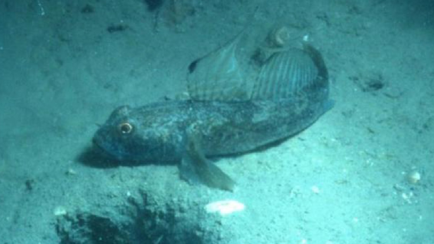
In the study, researchers from the University of Vienna applied high-resolution backscatter electron (BSE) imaging to otoliths from the black goby (Gobius niger), a common Adriatic fish. Unlike traditional light microscopy or secondary electron techniques, BSE exploits compositional contrasts in mineral density to visualize microstructures within the otolith matrix, including ultrafine growth rings.

“With the electron microscope, we were able to make even the smallest growth increments visible,” said lead author Isabella Leonhard, in a press release. “Typically, these rings form in a daily rhythm. In addition, micro-increments exist that form independently of the daily cycle – these sub-daily patterns reflect feeding, movement, environmental changes, or stressors the fish was exposed to.”

By fine-tuning scan settings and using extended acquisition times, the team detected up to 275 percent more microincrements in Holocene-aged otoliths than standard imaging methods allowed. Some bands were less than 0.5 µm wide – undetectable under light microscopy due to the opacity and diagenesis typical of ancient specimens. Crucially, preservation quality didn’t limit internal visibility: BSE revealed internal features even in otoliths over 7,600 years old.

The results offer a new path for sclerochronological studies of ancient fish, with potential applications in reconstructing environmental and ecological changes across millennia. “Our results show that fossil otoliths have enormous untapped potential,” said co-author Martin Zuschin. “They can help us to better understand the changes we are seeing today.”

The team recommends a combined workflow: LM screening for large datasets, with targeted BSE imaging of opaque or poorly preserved material. As otoliths from small, non-commercial species are rarely studied in such detail, the method also opens doors to long-term population studies beyond fisheries science.

Newsletters

Receive the latest analytical science news, personalities, education, and career development – weekly to your inbox.

Newsletter Signup Image

False

Advertisement

Recommended

False

Related Content

The Analytical Scientist Innovation Awards 2024: #3
Microscopy
The Analytical Scientist Innovation Awards 2024: #3

December 6, 2024

4 min read

Bruker’s multiphoton microscopy module, OptoVolt, ranks third in our Innovation Awards. Here, Jimmy Fong, product development lead, walks us through the major moments during development.

Let Me See That Brain
Microscopy
Let Me See That Brain

December 9, 2024

1 min read

TRISCO sets a new standard for 3D RNA imaging, delivering high-resolution and uniform images to offer insights into brain function and anatomy

Loading the Molecular Dice
Microscopy
Loading the Molecular Dice

December 12, 2024

2 min read

How a framework for controlling molecular reactions at the atomic scale has potential implications for nanotechnology, pharmaceutical synthesis, and clean energy research

Found in Translation
Microscopy
Found in Translation

December 16, 2024

1 min read

Cryo-EM addresses a key question in gene expression: how ribosomes initiate translation

False

The Analytical Scientist
Subscribe

About

  • About Us
  • Work at Conexiant Europe
  • Terms and Conditions
  • Privacy Policy
  • Advertise With Us
  • Contact Us

Copyright © 2025 Texere Publishing Limited (trading as Conexiant), with registered number 08113419 whose registered office is at Booths No. 1, Booths Park, Chelford Road, Knutsford, England, WA16 8GS.