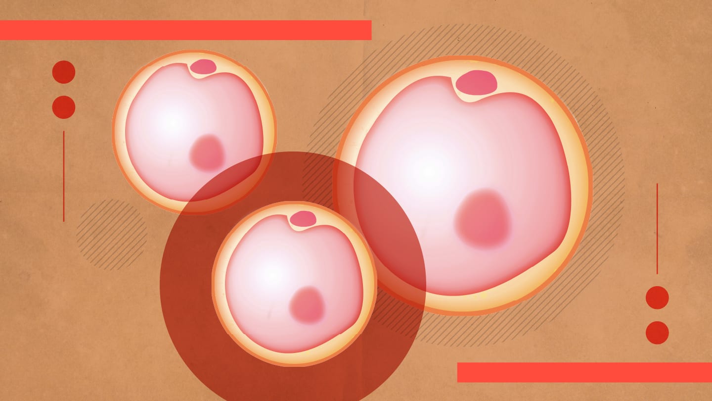
A study combining single-cell Raman spectroscopy with high-resolution imaging has mapped age-related metabolic changes in live mouse egg cells, offering noninvasive insight into why fertility declines with age.
The study was led by researchers at Peking Union Medical College Hospital and the University of Glasgow. Using 166 live mouse oocytes across developmental stages and ages – along with detailed spatial data from nine cells – the team traced molecular changes in lipid, protein, and cytochrome c content during oocyte maturation.
“Raman spectroscopy lets us capture a molecular fingerprint of living cells without labels or dyes,” said Jiabao Xu of the University of Glasgow, in the press release. By applying trajectory analysis to the spectral data, the team showed that although the nuclear maturation of older oocytes appeared intact, their metabolic profiles lagged behind – an uncoupling that points to developmental asynchrony.
One of the most notable findings was a dramatic loss of cytochrome c in aging oocytes. This key mitochondrial protein plays a central role in energy production, and its absence suggests mitochondrial dysfunction is a major driver of declining oocyte quality.
“These findings suggest that mitochondrial dysfunction is a central factor in the age-related decline of oocyte quality,” said co-author Hanbi Wang of Peking Union Medical College Hospital.
High-resolution Raman imaging further enabled the researchers to map the distribution of metabolites within individual oocytes. Older cells showed altered lipid and protein localization, supporting the idea of impaired energy metabolism and cellular remodeling.
Beyond basic research, the authors highlight the clinical potential of their method for fertility screening. “Our approach could eventually guide fertility treatments by identifying oocytes with the highest developmental potential,” Xu added.


